SA Node vs AV Node: 8 Key Differences in Heart Rhythm Control
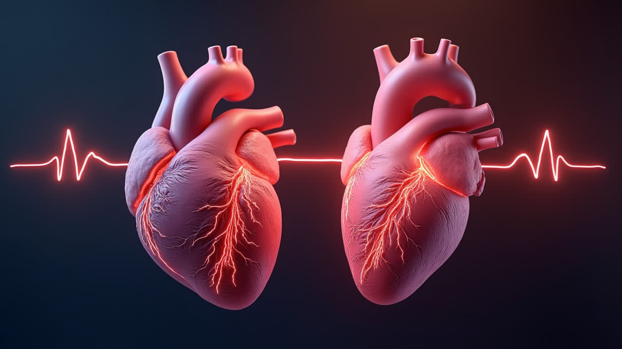
The heart's electrical system is a marvel of biological engineering. At its core are two specialized structures that control your heartbeat: the SA node and the AV node. Understanding the difference between these two cardiac components isn't just academic knowledge—it's essential for comprehending how your heart functions and what can go wrong in certain cardiac conditions.
Have you ever wondered what keeps your heart beating in its steady rhythm? Or why doctors pay special attention to these tiny structures during cardiac examinations? Today, we'll explore the fascinating world of cardiac conduction and unravel the key differences between these vital rhythm controllers.
The SA node (sinoatrial node) and AV node (atrioventricular node) form crucial parts of the heart's electrical conduction system. While they work together seamlessly in a healthy heart, they serve distinctly different functions that ensure your heart beats efficiently to circulate blood throughout your body. Let's dig deeper into what makes each node unique and how they contribute to the symphony of your heartbeat.
What is the SA Node?
The sinoatrial node, commonly known as the SA node, is a small, specialized tissue mass located in the right atrium of the heart. Specifically, it sits in the superior lateral wall, near where the superior vena cava enters the heart. This strategic position allows it to influence the entire cardiac muscle effectively.
Often called the "natural pacemaker" of the heart, the SA node generates electrical impulses that initiate each heartbeat. These impulses arise spontaneously due to the unique properties of the pacemaker cells that make up the node. Unlike other cardiac cells, pacemaker cells can depolarize automatically without needing external stimulation, creating a steady rhythm of electrical activity.
The SA node typically fires at a rate of 60-100 beats per minute in adults at rest, though this can vary significantly with factors like age, fitness level, and emotional state. When you exercise or experience stress, your sympathetic nervous system increases this firing rate, allowing your heart to pump more blood to meet increased oxygen demands. Conversely, during rest or sleep, your parasympathetic nervous system slows the SA node's firing rate, conserving energy when high cardiac output isn't needed.
Structurally, the SA node consists of specialized pacemaker cells surrounded by paranodal cells. These cells are smaller than typical atrial cells and contain fewer mitochondria. This unique cellular composition enables the SA node to fulfill its critical role as the primary initiator of cardiac contraction sequences.
What is the AV Node?
The atrioventricular node, or AV node, represents another key component in the heart's electrical conduction system. Located in the posterior septal wall of the right atrium, near the opening of the coronary sinus, this structure sits strategically at the boundary between the atria and ventricles.
Unlike the SA node, the AV node doesn't typically initiate electrical impulses under normal conditions. Instead, it serves as a critical relay station, receiving impulses from the SA node and transmitting them to the ventricles. This relay function might seem simple, but it's absolutely vital for proper cardiac function.
One of the AV node's most important roles is its ability to delay electrical impulses. When an impulse arrives from the SA node, the AV node holds it for approximately 0.09 seconds before passing it on to the ventricles. This brief delay serves a crucial physiological purpose—it allows the atria to complete their contraction and fully empty blood into the ventricles before ventricular contraction begins. Without this delay, the heart's pumping efficiency would be severely compromised.
The AV node also serves as a protective gatekeeper for your heart. If the atria begin firing excessively rapid impulses—as occurs in certain arrhythmias like atrial fibrillation—the AV node prevents all these impulses from reaching the ventricles. This protective function prevents the ventricles from beating dangerously fast, which could otherwise lead to insufficient filling time and reduced cardiac output.
In situations where the SA node fails, the AV node can act as a backup pacemaker, generating impulses at a rate of about 40-60 beats per minute. While this "junctional rhythm" is slower than the normal sinus rhythm, it provides a vital safety mechanism to maintain basic cardiac function until the primary pacemaker recovers or medical intervention occurs.
SA Node vs AV Node: Comparison Table
| Characteristic | SA Node | AV Node |
|---|---|---|
| Full Name | Sinoatrial Node | Atrioventricular Node |
| Primary Function | Generates cardiac impulses (primary pacemaker) | Relays and delays cardiac impulses |
| Location | Superior lateral wall of right atrium, near superior vena cava | Posterior septal wall of right atrium, near coronary sinus |
| Shape and Size | Longer, flattened, ellipsoidal | Short, half-oval shaped |
| Intrinsic Firing Rate | 60-100 beats per minute | 40-60 beats per minute |
| Transmission Path | Directly to right and left atria | To ventricles via bundle of His |
| Role in Cardiac System | Pacemaker (sets rhythm) | Pacesetter (controls contraction timing) |
| Regulatory Control | Autonomic nervous system | Primarily regulated by SA node |
Key Differences Between SA Node and AV Node
1. Function and Role
The most fundamental difference between these nodes lies in their primary functions. The SA node serves as the heart's natural pacemaker, spontaneously generating electrical impulses that initiate each heartbeat. In contrast, the AV node primarily functions as a relay station and electrical gatekeeper. It receives impulses from the SA node, delays them briefly, and then passes them on to the ventricles. This functional difference reflects their specialized roles in the cardiac conduction sequence.
2. Anatomical Location
Location differences are significant for understanding cardiac physiology. The SA node is positioned in the superior lateral wall of the right atrium, strategically placed near where the superior vena cava enters the heart. The AV node, however, sits at the junction between the atria and ventricles in the posterior septal wall of the right atrium, close to the opening of the coronary sinus. These distinct locations enable each node to perform its specialized function effectively.
3. Intrinsic Firing Rates
Another notable difference involves their intrinsic firing rates. The SA node naturally fires at 60-100 beats per minute in a resting adult, while the AV node has a slower intrinsic rate of only 40-60 beats per minute. Under normal conditions, the faster SA node overrides the AV node's rhythm. However, if the SA node fails, the AV node can take over pacemaking duties, albeit at a slower rate, in what's known as a junctional rhythm.
4. Impulse Transmission Pathways
The nodes differ significantly in how they transmit electrical impulses. The SA node sends signals directly to the atrial myocardium, causing both atria to contract almost simultaneously. In contrast, the AV node transmits impulses to the ventricles through specialized conduction pathways—the bundle of His, bundle branches, and Purkinje fibers. This organized pathway ensures coordinated ventricular contraction from apex to base, maximizing cardiac efficiency.
5. Regulatory Mechanisms
The regulation of these nodes also differs considerably. The SA node is directly regulated by both branches of the autonomic nervous system—sympathetic stimulation increases its firing rate, while parasympathetic stimulation decreases it. The AV node, however, is primarily regulated by the SA node itself. While it does receive autonomic input, its activity is largely dependent on the signals it receives from the SA node upstream in the conduction pathway.
6. Cellular Composition
At the cellular level, both nodes consist of specialized cardiac cells, but their compositions differ. The SA node contains a higher proportion of P cells (primary pacemaker cells) with fewer contractile elements and more sarcoplasmic reticulum. The AV node contains more transitional cells and fewer pure pacemaker cells, reflecting its primary role in conduction rather than impulse generation.
Similarities Between SA Node and AV Node
Despite their differences, these cardiac structures share several important similarities worth noting:
- Both are specialized conduction tissues located in the right atrium of the heart
- Both consist of modified cardiac muscle cells with specialized electrical properties
- Both play essential roles in maintaining normal cardiac rhythm and function
- Both can act as pacemakers, though the SA node normally dominates
- Both receive input from the autonomic nervous system, though to different degrees
- Both can be affected by similar cardiac medications, particularly those that affect ion channels
These shared characteristics highlight how these structures evolved as specialized components of a unified cardiac conduction system. Working together, they ensure that your heart beats in a coordinated, efficient manner to maintain circulation throughout your body.
Clinical Significance of SA and AV Nodes
Understanding the differences between these nodes has profound clinical importance. Disorders affecting either node can lead to various cardiac arrhythmias with potentially serious consequences. For instance, dysfunction of the SA node can cause sick sinus syndrome, characterized by inappropriate heart rates and rhythm disturbances. Similarly, AV node dysfunction may result in heart blocks, where impulse transmission between atria and ventricles becomes delayed or interrupted.
Many cardiac medications target these nodes to treat arrhythmias. Beta-blockers and calcium channel blockers can slow conduction through both nodes, helping control rapid heart rates. In some cases, when medication proves ineffective, procedures like catheter ablation may modify the conduction properties of these nodes to treat certain arrhythmias.
Modern cardiac pacemakers are often implanted when these natural pacemakers fail. Understanding the normal function of the SA and AV nodes helps physicians program these devices to mimic natural cardiac rhythm as closely as possible. Some advanced pacemakers even have separate leads for the atria and ventricles, preserving the natural delay between atrial and ventricular contraction that the AV node normally provides.
Conclusion
The SA node and AV node represent specialized components of the heart's electrical conduction system, each with distinct functions, properties, and locations. The SA node, as the primary pacemaker, initiates the heartbeat by generating electrical impulses at a rate of 60-100 beats per minute. The AV node serves as both a relay station and a gatekeeper, delaying impulses briefly to allow proper coordination between atrial and ventricular contraction.
These differences aren't merely academic distinctions. They represent evolutionary adaptations that ensure the heart functions with maximum efficiency as a pumping organ. The specialized roles of these nodes allow for the precise timing of cardiac chamber contractions, optimizing blood flow and ensuring adequate circulation throughout the body.
By understanding how these nodes differ and work together, we gain deeper insights into both normal cardiac physiology and the mechanisms underlying various cardiac arrhythmias. This knowledge continues to inform both pharmacological approaches to treating heart rhythm disorders and the development of increasingly sophisticated cardiac devices that can support or replace natural pacemaker function when needed.
Frequently Asked Questions
Can the heart function if the SA node stops working?
Yes, the heart can continue functioning even if the SA node fails, though typically at a slower rate. When the SA node stops working properly, other parts of the cardiac conduction system can take over pacemaking duties. The AV node usually assumes this role first, generating impulses at a rate of 40-60 beats per minute (compared to the SA node's 60-100 beats per minute). If both the SA and AV nodes fail, specialized cells in the ventricles can generate impulses at about 20-40 beats per minute. This hierarchical backup system helps ensure that the heart continues beating even when its primary pacemaker malfunctions, though medical intervention is often necessary to restore optimal function.
How do medications affect the SA and AV nodes differently?
Various cardiac medications can affect the SA and AV nodes differently based on their specific mechanisms and the nodes' unique properties. Beta-blockers and calcium channel blockers can slow conduction through both nodes, but often affect the SA node more significantly, reducing heart rate by decreasing its automaticity. Digoxin predominantly affects the AV node, increasing its refractory period and slowing conduction, which is particularly useful in controlling ventricular rate in atrial fibrillation. Atropine blocks parasympathetic effects, primarily increasing SA node firing rate while having less impact on the AV node. These differential effects allow physicians to target specific aspects of cardiac conduction when treating various arrhythmias.
What happens during different types of heart block involving the AV node?
Heart blocks occur when there's interference with impulse transmission through the AV node, and they're classified by severity. In first-degree AV block, impulses are delayed but all eventually reach the ventricles, appearing as prolonged PR intervals on an ECG. Second-degree blocks come in two types: Mobitz type I (Wenckebach), where PR intervals progressively lengthen until a beat is dropped, and Mobitz type II, where some atrial impulses fail to reach the ventricles without warning. The most severe form, third-degree or complete heart block, occurs when no impulses pass through the AV node, causing the atria and ventricles to beat independently. This complete disconnection often requires immediate intervention with a temporary or permanent pacemaker to maintain adequate circulation.